Knowing is better than not knowing.
Especially when it comes to breast cancer.
Suppress breast fat in MRI imaging with CaliberMRI’s
unique fat mimic, developed in collaboration with the
University of California, San Francisco and NIST.
Standardize ADC and T1 relaxation values with
CaliberMRI’s human tissue mimic solutions developed in
over a decade of collaboration with leading qMRI
researchers and scientists.
Expand the temperature range capabilities of MRI standardization with the one-of-a-kind MR-readable thermometer
MRI standardization just got even easier with the integration of the CMRI patented LC MR-Readable Thermometer, developed in collaboration with NIST.1 Now qMRI advancers can measure and directly observe the phantom temperature between 15°C and 24°C.
1 | This product is covered by United States patent number 10,809,331.
Know the specs
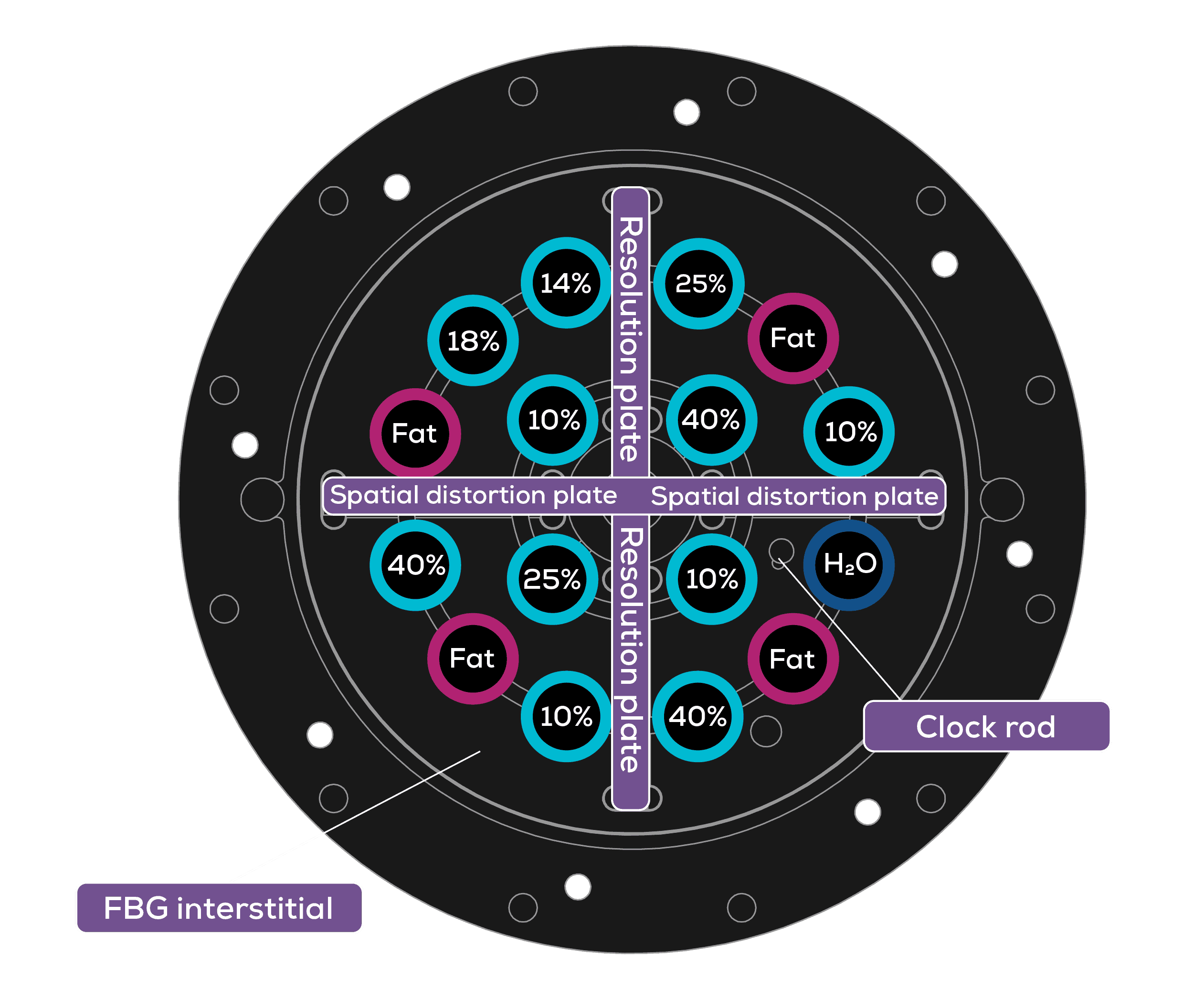
The Contrast and Diffusion sides are encased in robust, semi-rigid double-walled shells. Between the walls is a fat mimic designed for comprehensive fat suppression assessment.
Evaluated MRI Characteristics
- B1 and B0 non-uniformity, geometric linearity, gradient amplitude
- System center frequency drift (short time duration)
- Resolution and SNR
- Accuracy and precision of T1 measurements
- Accuracy and precision of the Apparent Diffusion Coefficient (ADC)
The Contrast Side for T1 Relaxation Standardization
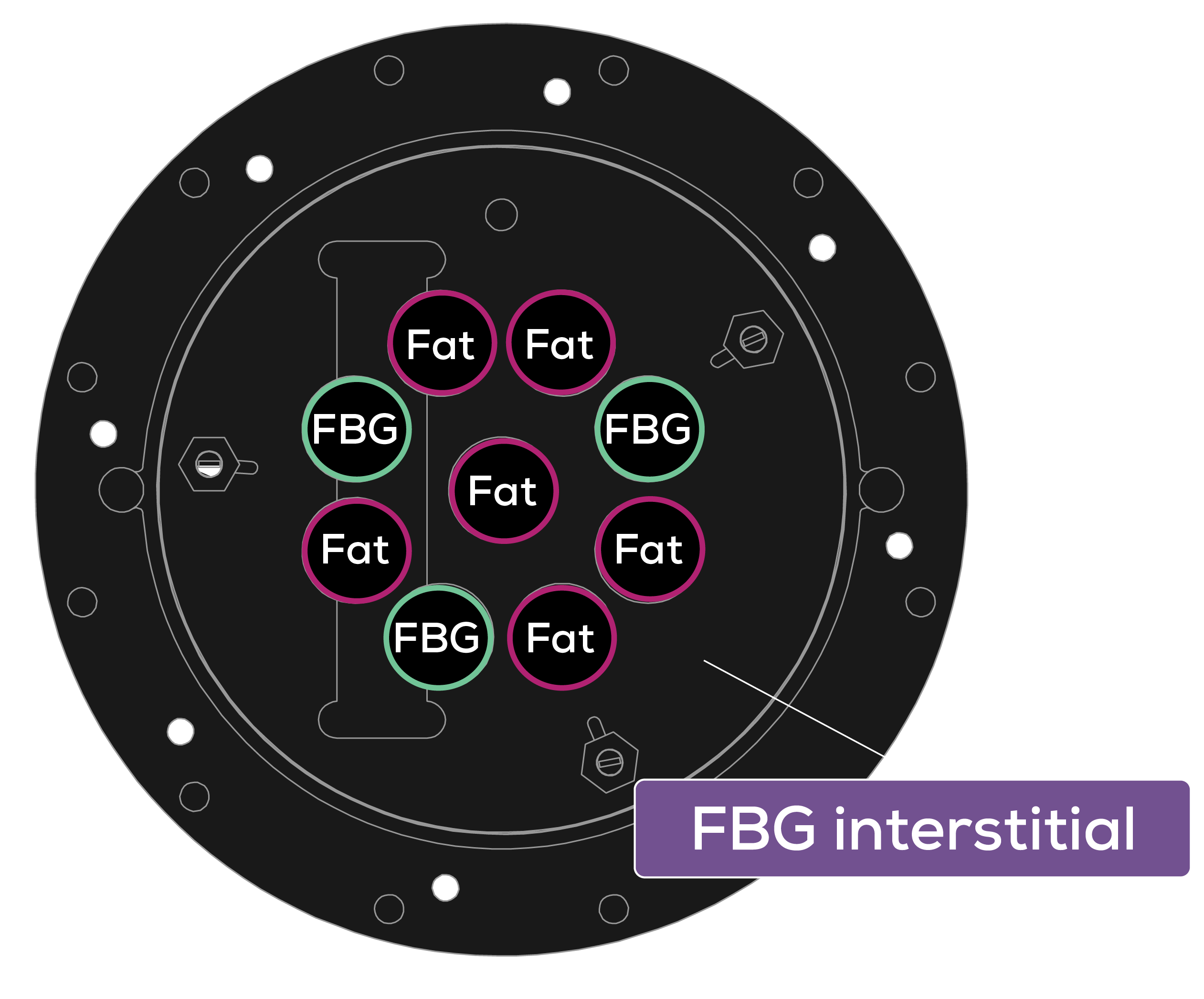
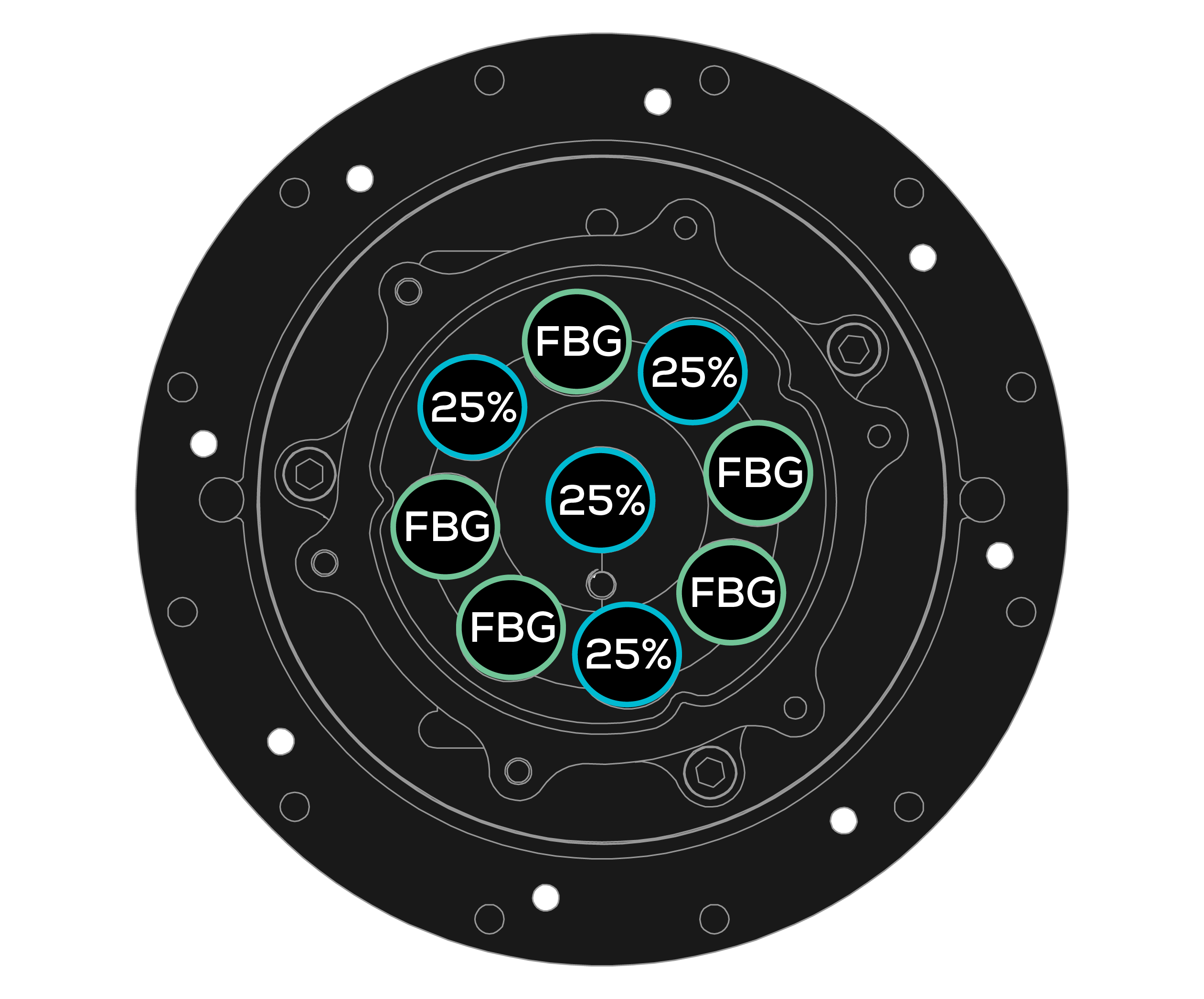
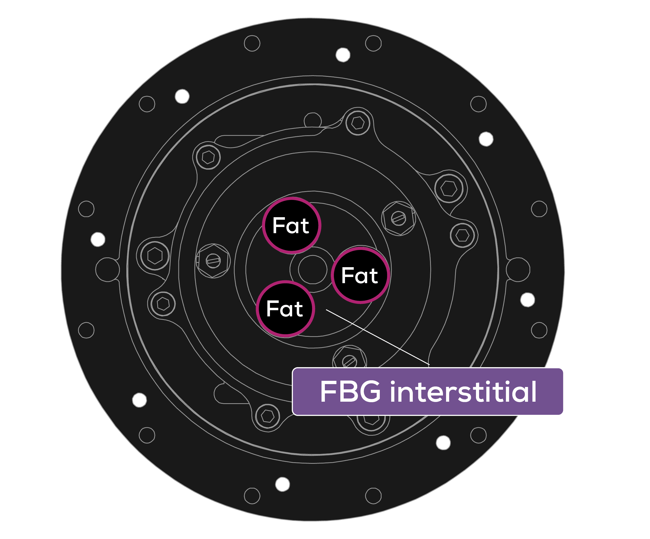
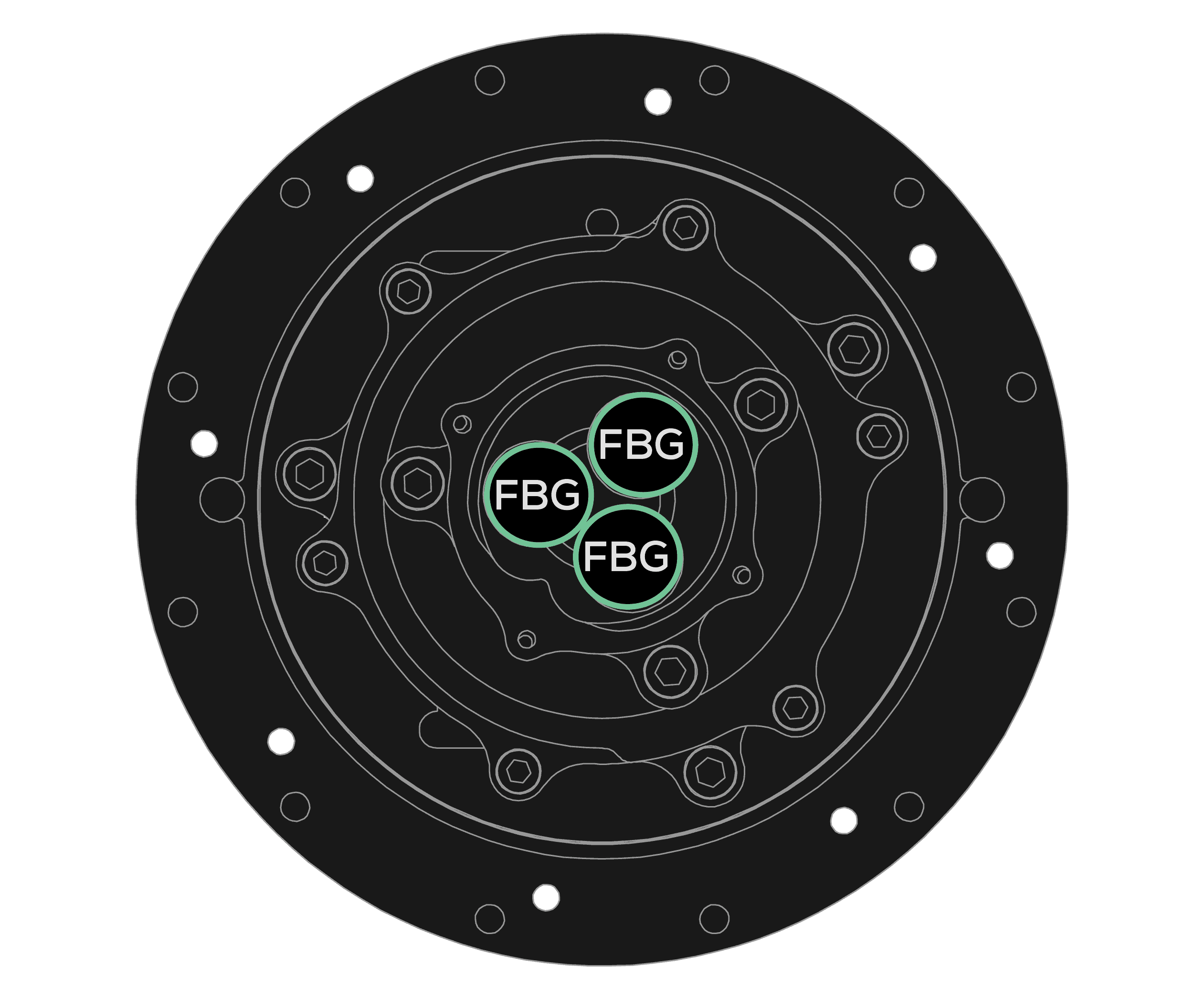
The Contrast Side contains an array of fat, dense fibroglandular (FBG) tissue, and PVP mimics that aid in fat suppression, and T1 mapping and standardizing.
- 24 contrast spheres for fat T1 (335 ms, 3.0 T at 20°C) and FBG dense tissue T1 (1580 ms,
3.0 T at 20°C) standardization.
Automate Breast MRI QC from A to Z with
CaliberMRI’s qCal-MR Software
Compare your MRI measurement outputs with NIST-traceable values and
share studies with your qMRI research colleagues around the world.

